Journal of Cardiovascular Medicine and Cardiology
Pulmonary Artery Aneurysm - Report of a Case and Review of Literature
Author and article information
Cite this as
Sargur R, Sudarshana Murthy KA (2015) Pulmonary Artery Aneurysm - Report of a Case and Review of Literature. J Cardiovasc Med Cardiol 2(1): 029-031. 10.17352/2455-2976.000014
Copyright License
© 2015 Sargur R, et al. This is an open-access article distributed under the terms of the Creative Commons Attribution License, which permits unrestricted use, distribution, and reproduction in any medium, provided the original author and source are credited.Abstract
Pulmonary artery aneurysm is a very rare condition. Clinical experience is limited and current knowledge is mainly derived from autopsy findings, however, Pulmonary arterial aneurysms are being detected more frequently with modern techniques of echocardiography and angiography. We report a case of pulmonary artery aneurysm presenting with pulmonary hypertension. We will present a review of literature and discuss the differential diagnosis, presentation and management.
Case Report
A 57 year old female was admitted with an acute presentation of increasing breathlessness, pain in interscapular area associated with dizziness and vomiting. There was no orthopnea, paroxysmal nocturnal dyspnea or chest pain.
Past medical history was significant for recurrent SVT between the ages of 15 and 30 which she was able to control by vagal manoeuvres. She was diagnosed to have pulmonary hypertension but no cause was found. She had progressive dyspnoea for last 5 years and was admitted with cardiac failure on 3 occasions.
She was tachycardic with a heart rate of 100 /m and tachypnec with a respiratory rate of 30/m, normotensive -130/90 - no difference in blood pressures in right and left arm, there was no peripheral edema. Examination of precardium revealed RVH with pulmonary hypertension with a diffuse apical impulse, parasternal heave, RVS3, EDM in pulmonary area and parasternal area. There was a dull note in left infra-axillary and infra-scapular areas with absent breath sounds suggestive of a pleural effusion. Her haemoglobin was low at 8 g/l.
ECG showed sinus tachycardia and right ventricular dominance. Chest x-ray confirmed the presence of a pleural effusion (Figure 1) which on aspiration was confirmed to be a haemo-thorax.
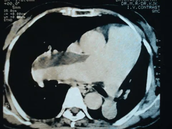
A CT thorax with contrast showed enlarged main pulmonary artery, right and left pulmonary arteries with a thrombotic fusiform aneurysm of right Pulmonary artery. There was a non enhancing thrombus progressively reducing the lumen of the LPA (Figure 2). A diagnosis of aneurysm of PA was made. Despite intensive management, unfortunately, she deteriorated over the next 24 hours and died.
Discussion
Aneurysm of pulmonary arterial trunk or of its major branches is rare. Most involve the main trunk of the Pulmonary artery with or without involvement of its branches.
Frequently aneurysms of the PA are associated with or result from congenital cardiac defects.
Pulmonary arterial aneurysms are usually associated with congenital heart anomalies, infection, collagen vascular diseases or degenerative changes of the elastic media [1] (Table 1). Most cases are diagnosed at autopsy, but more and more cases are being diagnosed incidentally by imaging modalities [2-6] of which a number of cases have been successfully treated by surgery.
Table 1:
Causes of Pulmonary Arterial Aneurysm.
Abbreviations: MPA: Main Pulmonary Artery; RPA: Right Pulmonary Artery; RPA: Right Pulmonary Artery
In our patient, there were no history and clinical features suggestive of coexisting congenital heart disease, stigmata of connective tissue disease and Marfan's Syndrome. Tests for tuberculosis and syphilis was negative. There was no evidence of vegetations on echocardiography.
The majority of cases arise from the proximal pulmonary arteries and compress the surrounding parenchyma and vasculature [1,7]. The clinical manifestations depend on the cause and the location and size of the aneurysm. The right ventricle is frequently enlarged. Although usually asymptomatic, they may cause dyspnoea, cough, haemoptysis, and chest pain. Cardiac failure and rupture are the commonest causes of death. An increasing number have been successfully treated with surgery in recent years, however, massive and often fatal haemoptysis reportedly occurs in 20-60% of cases, particularly with the solitary peripheral lesions [8].
Our patient had moderate to severe pulmonary Hypertension. Severe pulmonary hypertension has been reported in pulmonary arterial aneurysm with persistent ductus arteriosus [11]. Primary Pulmonary hypertension may lead to fusiform dilatation of the pulmonary arteries but aneurysmic dilation is rare. Pulmonary hypertension secondary to repeated pulmonary emboli is the most likely cause in our patient.
The differential in our patient is one of a mild occult congenital cardiac defect leading to severe pulmonary arterial dilatation and aneurysm complicated with pulmonary hypertension secondary to pulmonary embolisation. The second likely possibility would be one of idiopathic dilatation of the pulmonary artery.
Turano and Gambaccini [12] reported raised pulmonary arterial pressures in patients with dilated central pulmonary arteries but it is debatable whether what they were seeing was changes secondary to chronic pulmonary hypertension.
Pulmonary artery aneurysms in adults is a very rare entity and there are no clear guidelines for optimal treatment. Operative treatment is recommended for patients with a risk of rupture, which is not well defined. Most of the evidence comes from case reports and case series.
Conservative treatment has been advocated when there was no left-to-right intracardiac shunt or significant pulmonary arterial hypertension, which resulted in a relatively benign prognosis with an uncomplicated course after 1 to 7 years of follow-up [13]. Surgical options include aneurysmorrhaphy, replacement with a Dacron graft or pulmonary allograft, and combined use of a stentless bioprosthesis and a Dacron prosthesis [14-16].
Our patient would have had better outcome if a diagnosis of pulmonary arterial aneurysm was made earlier. This case report highlights the need for proactive investigation of aetiology of pulmonary hypertension in all cases.
Acknowledgement
Manuscript was prepared and reviewed by Dr. Sargur and Dr. Murthy.
References
- Trell E (1973) Pulmonary arterial aneurysm. Thoraxx 28: 644-649.
- Wu WS (2003) Images in cardiovascular medicine. Huge calcified pulmonary arterial aneurysm. Circulation 107: 2280-2281.
- Janssens F, Verswijvel G, Colla P, Smits J, Gubbelmans H, et al. (2003) Proximal pulmonary artery aneurysm. JBR-BTR 86: 83-85.
- Khalil A, Parrot A, Fartoukh M, Marsault C, Carette MF (2006) Images in cardiovascular medicine. Large pulmonary artery aneurysm rupture in Hughes-Stovin syndrome: multidetector computed tomography pattern and endovascular treatment. Circulation 114: e380- e381.
- Hartshorne MF, Eisenberg B (1989) CT diagnosis of a giant central pulmonary artery aneurysm arising quickly after pulmonary embolic disease. AJR Am J Roentgenol 153: 190-191.
- Cherwek H, Amundson S (2003) Images in clinical medicine. Pulmonary-artery aneurysm. N Engl J Med 348: e1.
- Gibbs PM, Hami A (1995) Pulmonary arterial aneurysm presenting as an endobronchial mass. Thorax 50: 1013-1014.
- Ungaro R, Saab S, Almond CH, Kumar S (1976) Solitary peripheral pulmonary artery aneurysms. Pathogenesis and surgical treatment. J Thorac Cardiovasc Surg 71: 566-571.
- Yakut ZI, Odev K (2007) Pulmonary and cardiac involvement in Behçet disease: 3 case reports.Clin Appl Thromb Hemost 13: 318-322.
- Best J (1967) Dissecting aneurysm of the pulmonary artery with multiple cardiovascular abnormalities and pulmonary hypertension. Med J Aust 2: 1129-1130.
- Ravines HT (1960) Dissecting hematomas of intrapulmonary arteries in a case of pulmonary hypertension associated with patent ductus arteriosus. J Thorac Cardiovasc Surg 39: 760-766.
- Turano L, Gambaccini P (1958) Anatomical and roentgenological aspects of the solitary s.c. idiopathic dilatation of the pulmonary artery (with some remarks regarding its pathogenesis). Sci Med Ital 6: 435-462.
- Tami LF, McElderry MW (1994) Pulmonary artery aneurysm due to severe congenital pulmonic stenosis. Case report and literature review. Angiology 45: 383-390.
- Agarwal S, Chowdhury UK, Saxena A, Ray R, Sharma S, et al. (2002) Idiopathic Pulmonary Aneurysm. Asian Cardiovasc Thorac Ann 10: 167-169.
- Garcia-Rinaldi R, Howell JF (1976) Aneurysm of the main pulmonary artery: long-term survival after aneurys-morrhaphy and closure of a ventricular septal defect. Ann Thorac Surg 21: 180-183.
- Casselman F, Deferm H, Peeters P, Vanermen H (1995) Aneurysm of the left pulmonary artery: surgical allograft repair. Ann Thorac Surg 60: 1423-1425.
Advertisement






Figure 1
Chest X-ray showing pleral effusion and prominent Pulmonary arterial shadow.
Figure 1: Chest X-ray showing pleral effusion and prominent Pulmonary arterial shadow.