Journal of Cardiovascular Medicine and Cardiology
Pathological Biomineralization in the Calcific Aortic Valve
Author and article information
Cite this as
Cavarretta E, Maras A (2016) Pathological Biomineralization in the Calcific Aortic Valve. J Cardiovasc Med Cardiol 3(1): 045-046. DOI: 10.17352/2455-2976.000032
Copyright License
© 2016 Cavarretta E,et al. This is an open-access article distributed under the terms of the Creative Commons Attribution License, which permits unrestricted use, distribution, and reproduction in any medium, provided the original author and source are credited.Abstract
Background: Pericardial tamponade, a life-threatening condition caused by the accumulation of fluid in the pericardial sac, can be acute or chronic. Mortality and morbidity can be minimized on prompt diagnosis and treatment with percutaneous drainage.
Materials and methods: 246 patients admitted with cardiac tamponade between Jan 2010 and Aug 2014 was enrolled in the study. After clinical examination and echocardiographic assessment, all the patients were subjected to emergency pericardiocentesis (both diagnostic and therapeutic). Three samples of the aspirate were sent for biochemical, serological, and cytological analysis. The data collected was then analyzed.
Results: Tuberculosis is the commonest cause in our study. Post procedural and post-surgical causes are insignificant in numbers. Overall mortality rate in our study is 6% which is much lesser than other studies. Procedure related cardiac tamponade mortality rate was 6%.
Conclusion: Initial assessment with clinical, serologic, and echocardiographic investigation and careful follow-up can in most cases yield a causal diagnosis. The prognosis depends chiefly upon the patient’s underlying disease.
Editorial
The prevalence of moderate to severe calcific aortic valve stenosis in patients ≥75 years old is 2.8% and only 40% of patients with surgical indication undergo aortic valve replacement because of high perioperative risk, older age, lack of symptoms, and patient/family refusal [1]. In the absence of hemodynamically significant left ventricular (LV) outflow obstruction, calcific aortic valve disease (CAVD) prevalence raises up to 25% in patients aged from 65 to 74 years old [2,3] and independently predicts cardiovascular (CV) event, overall and CV mortality. As the population ages and CAVD incidence and prevalence increase, it is crucial a deeper understanding of the patho-physiology of heart valve calcification that could provide novel insight into medical therapeutic approaches to delay or modify the disease course. In the human body, several physiological processes of calcification take places and mineralized deposits are present, as bones, enamel and dentin. More than that, pathological mineralization can lead to ectopic calcification and pathologies as urinary stones, vascular calcification and calcific heart valve stenosis. Macroscopically, in aortic valve sclerosis there is an initial thickening of the valve leaflets and formation of calcium nodules, usually corresponding to the nodules of Arantio near the aortic surface, in association to angiogenesis, while end-stage calcific aortic stenosis is characterized by large, heavily calcific, nodular masses within the aortic cusps that protrude into the sinuses of Valsalva, thus interfering with valve opening along the aortic surface. While in the past heart valve calcification was seen as a passive, degenerative course of aging, evidences have shown that is an active, cell-mediated process with similarities to bone development (osseus metaplasia) [4]. In fact all of the markers of bone differentiation including the non-collagenous bone matrix proteins osteopontin, osteocalcin, bone sialoprotein and the osteoblast transcription factor Cbfa1 were found increased in the calcific aortic valve that express an osteoblast phenotype [5]. Studies on the pathobiology of CAVD showed that the calcific process begins deep into the valve tissue, near the margins of attachment. As for vascular calcification, oxidized low-density lipoproteins (LDL) play an important role in the initiation [6], in association with angiotensin-converting enzyme (ACE) and its product, angiotensin II. ACE has been found colocalized with apolipoprotein B, the primary protein found on LDL, in aortic valve lesions and also associated with plasma LDL, which may deliver ACE to aortic valve lesions [7]. In addition, oxidative stress is increased in calcified aortic valves, due to reduction in expression and activity of antioxidant enzymes and perhaps to uncoupled nitric oxide synthases (NOS) activity [8]. The presence of reactive oxygen species associated to inflammatory cytokines, growth factors, altered shear stress and hypertension could be the triggers for valvular interstitial cells (VICs) pathological differentiation in myofibroblasts and osteoblasts. The so activated VICs produce matrix metalloproteinases, involved in extracellular matrix remodeling, and increase angiogenic activity, thus creating the perfect microenvironment to promote mineralization. In particular valve calcification is composed of calcium phosphate crystals that form biominerals, deposited on a bone-like matrix of collagen, osteopontin, and other minor bone matrix proteins. The spatial organization of fibrils can create the crystal-growing niche, where pathological biomineralization can take place because of the specific physical-chemical characteristics that lead to mineral precipitation so that crystal can grow with a regular shape [9]. Nowadays bioapatite is the term most used in different scientific fields to indicate the mineral component of normal mineralized tissues and pathological calcification. This term is considered as indicative of an impure carbonate-containing apatite but, at present, the International Mineralogical Association Commission on New Minerals, Nomenclature and Classification (IMA-CNMNC) has not fully accepted it. We used infrared and Raman spectroscopy to demonstrate the exact crystallographic structure of bioapatite. In fact, in the calcified deposits, after calcium [Ca] and phosphorus [P], the carbonate group [CO3] is the most important constituent. The percentage of [CO3] ranges from 5 to 10% in weight and its dominant location is in place of [PO4] [10]. The [CO3] substitution is linked to the major solubility and chemical reactivity of the inorganic phase both in vitro and in vivo. Moreover, nanometer scale investigations revealed that the pathological crystals are closely bound with the organic matrix and are characterized by variable size and shape, organized on different spatial scale. There is a high heterogeneity within calcific aortic valve nodules, where fully mineralized and partially mineralized areas are both present, representing two different stages of pathological mineralization [11]. Moreover, slightly but significant differences in terms of Ca and P amount exist between tricuspid and bicuspid CAVD. Even if macroscopically bicuspid valves show an accelerated and heavier calcification, chemically the concentration of Ca:P and their the mean weight % are significantly reduced compared to tricuspid aortic valve or calcific mitral valve (Figure 1).
Our nanoscale observations indicate that the formation of pathological bioapatite nanocrystals within heart valves is related to the presence of a highly heterogeneous bioapatite. Growth processes may occur in different microenvironments (each one with its own physico-chemical characteristics) influencing the shape of the biomineralization.
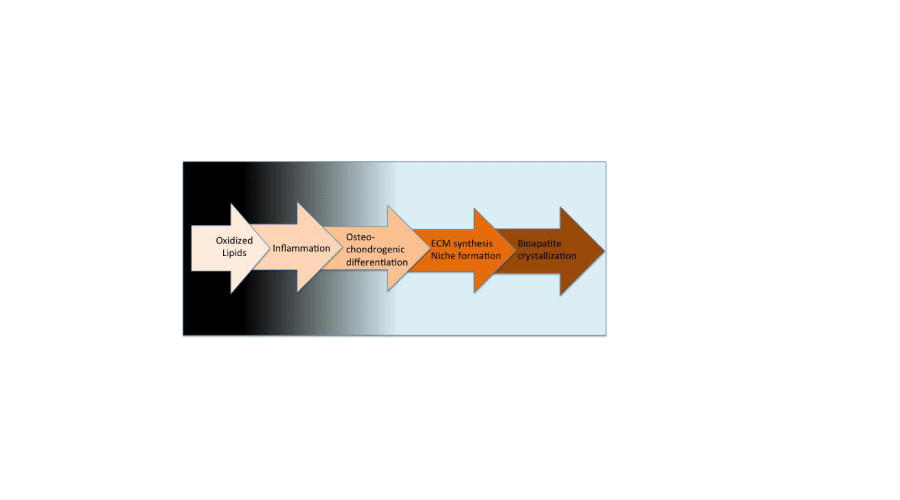
The presence of compartmental niches within the extracellular matrix assumes a relevant role in the formation of ectopic biomineralization in human heart valves as well as the action of the organic substrate on the crystals features.
These innovative findings at nanometer scale unveil novel insight into pathological calcification and could open new perspective to promote further studies on a novel medical approach to treat aortic valve calcification.
References
- Mozaffarian D, Benjamin EJ, Go AS, Arnett DK, Blaha MJ, et al. (2016) Heart Disease and Stroke Statistics-2016 Update: A Report From the American Heart Association. Circulation 133 :e38-60 .
- Völzke H, Haring R, Lorbeer R, Wallaschofski H, Reffelmann T, et al. (2010) Heart valve sclerosis predicts all-cause and cardiovascular mortality. Atherosclerosis 209: 606-610 .
- Owens DS, Budoff MJ, Katz R, Takasu J, Shavelle DM, et al. (2012) Aortic valve calcium independently predicts coronary and cardiovascular events in a primary prevention population. JACC Cardiovasc Imaging 5: 619-625 .
- Rajamannan NM, Evans FJ, Aikawa E, Grande-Allen KJ, Demer LL, et al. (2011) Calcific aortic valve disease: not simply a degenerative process: A review and agenda for research from the National Heart and Lung and Blood Institute Aortic Stenosis Working Group. Executive summary: Calcific aortic valve disease-2011 update. Circulation 124: 1783-1791 .
- Caira FC, Stock SR, Gleason TG, McGee EC, Huang J, et al. (2006) Human degenerative valve disease is associated with up-regulation of low-density lipoprotein receptor-related protein 5 receptor-mediated bone formation. J Am Coll Cardiol 47: 1707-1712 .
- Olsson M, Thyberg J, Nilsson J (1999) Presence of oxidized low density lipoprotein in nonrheumatic stenotic aortic valves. Arterioscler Thromb Vasc Biol 19: 1218-1222 .
- O'Brien KD, Shavelle DM, Caulfield MT, McDonald TO, Olin-Lewis K, et al. (2002) Association of angiotensin-converting enzyme with low-density lipoprotein in aortic valvular lesions and in human plasma. Circulation 106: 2224-2230 .
- Miller JD, Chu Y, Brooks RM, Richenbacher WE, Peña-Silva R, et al. (2008) Dysregulation of antioxidant mechanisms contributes to increased oxidative stress in calcific aortic valvular stenosis in humans. J Am Coll Cardiol 52: 843-850 .
- Cottignoli V, Relucenti M, Agrosì G, Cavarretta E, Familiari G, et al. (2015) Biological Niches within Human Calcified Aortic Valves: Towards Understanding of the Pathological Biomineralization Process. Biomed Res Int 2015: 542687 .
- Mangialardo S, Cottignoli V, Cavarretta E, Salvador L, Postorino P, et al. (2012) Pathological biominerals: Raman and infrared studies of bioapatite deposits in human heart valves. Appl Spectrosc 66: 1121-117 .
- Cottignoli V, Cavarretta E, Salvador L, Valfré C, Maras A (2015) Morphological and chemical study of pathological deposits in human aortic and mitral valve stenosis: a biomineralogical contribution. Patholog Res Int 2015: 342984 .
Advertisement