Journal of Cardiovascular Medicine and Cardiology
Large Thrombotic Coronary Artery Aneurysm Treated With a Covered Stent Using OCT for Procedural Guidance
Author and article information
Cite this as
Ferre GF, Galache Osuna JG, de Miguel JAD, Sánchez Rubio J (2017) Large Thrombotic Coronary Artery Aneurysm Treated With a Covered Stent Using OCT for Procedural Guidance. J Cardiovasc Med Cardiol 4(2): 023-024. 10.17352/2455-2976.000043
Copyright License
© 2017 Ferre GF, et al. This is an open-access article distributed under the terms of the Creative Commons Attribution License, which permits unrestricted use, distribution, and reproduction in any medium, provided the original author and source are credited.Abstract
Background: The study of coronary artery disease has been quite thorough. But there is few study on coronary vein disease. In clinical practice, some patients have suspicious symptoms of coronary artery disease without any evidence of coronary artery spasm, but coronary angiography showed no obvious stenosis in coronary artery. It may have relationship to coronary venous dysfunction. This study aims to develop a coronary vein ligation model, to investigate the effect of coronary vein occlusion on myocardial cells in normal miniature pigs.
Methods: Adult minifies were divided into control group and intervention group. The coronary venous occlusion model was made by clamping the central vein. Myocardium was isolated from the myocardium and the myocardium was observed by transmission electron microscopy.
Results: There were no significant changes in the sham operation group (control group). Mitochondrial edema, cristae broken, hyperplasia and auto phage some appeared in the operation group (intervention group).
Conclusion: The ligation of the coronary vein causes dysfunction of myocardial cells. It gives us inspiration, coronary vein occlusive disease may be underestimated. It may be some causes of angina for patients with normal coronary angiography.
Letter to Editor
Coronary artery aneurysm (CAA) is an uncommon finding in patients undergoing a coronary angiography, with an incidence of less than 5% [1]. CAA is defined as coronary dilation of more than 1,5 times the adjacent vessel diameter. The majority of CAA in adults are of an atherosclerotic origin [2].
Treatment of CAA remains challenging. In the past years surgery was the only successful approach of symptomatic CAA associated with coronary artery stenosis. Lately a less aggressive strategy has emerged with the inclusion of covered stents (CS). However the number of reported cases of CS in the treatment of CAA is low.
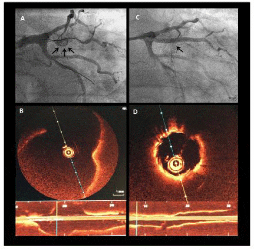
We present a case of a 79 years old man admitted to our hospital for a non ST-segment elevation acute myocardial infarction. The coronary angiogram revealed a large CAA at the proximal portion of the ramus intermedius followed by a severe stenosis. There was a large amount of intraluminal thrombus within the aneurysm (Figure 1A). In order to treat the coronary stenosis and exclude the large thrombotic CAA it was decided to use a CS. Optical coherence tomography (OCT) was performed before percutaneous coronary intervention (PCI) which was useful to choose stent dimension (Figure 1B). A 3x26mm polyurethane Papyrus stent (Biotronik, Berlin, Germany) was positioned in the proximal intermedius vessel after balloon predilation (Figure 1C). No distal embolization occurred. The final angiogram and OCT demonstrated the complete exclusion of the CAA as well as stenosis elimination (Figure 1D). The patient was discharged with the association of Acetylsalicylic acid and Ticagrelor, no anticoagulation was prescribed.
Covered stents have been classically used to seal coronary perforations and vein grafts [3]. It was proposed that the CS technology could be useful to seal the vessel lumen of coronary aneurysms. In this case of a large CAA complicated with an acute myocardial infarction the Papyrus CS demonstrated to be feasible and safe. The optimal imaging of OCT could provide a more accurate measurement of the CAA and vessel margins helping us to choose the appropriate stent. Despite initial excellent result a close follow up is needed because of the increased risk of restenosis and thrombosis described in these types of stents [4].
References
- Swaye PS, Fisher LD, Litwin P, Vignola PA, Judkins MP, et al. (1983) Aneurysmal coronary artery disease. Circulation 67: 134-138. Link: https://goo.gl/rpdoKA
- Syed M, Lesch M (1997) Coronary artery aneurysm: a review. Prog Cardiovasc Dis 40: 77-84. Link: https://goo.gl/COkmyd
- Elsner M, Auch-Schwelk W, Britten M, Walter DH, Schächinger V, et al. (1999) Coronary stent grafts covered by a polytetrafluoroenthylene membrane. Am J Cardiol 84: 335-338. Link: https://goo.gl/e6rO6R
- Briguori C, Sarais C, Sivieri G, Takagi T, Di Mario C, et al. (2002) Polytetrafluoroethylene-covered stent and coronary artery aneurysm. Catheter Cardiovasc Interv 55: 326-330. Link: https://goo.gl/irvmEY
Advertisement