Journal of Cardiovascular Medicine and Cardiology
Perforation of a side branch of coronary artery during coronary angiography: A rare complication
Bhupendra Kumar Sihag1, Pruthvi CR2 and Krishna Prasad3*
2Senior Resident, Department of Cardiology, Post Graduate Institute of Medical Education and Research, Chandigarh, India
3Senior Resident, Cardiology, Post Graduate Institute of Medical Education and Research, India
Cite this as
Sihag BK, Pruthvi CR, Prasad K (2020) Perforation of a side branch of coronary artery during coronary angiography: A rare complication. J Cardiovasc Med Cardiol 7(2): 136-137. DOI: 10.17352/2455-2976.000128Coronary angiography in current day scenario is a very safe procedure. Complications are very rare occurring in less than 1 % of cases. We report a case of coronary artery side branch perforation during angiography. Injection of contrast into a side branch following accidental super selective intubation, lead to the perforation at the tip and contrast extravasation. Patient was managed conservatively as he did not develop any hemodynamic compromise or pericardial effusion. Check angiography showed no leak from the perforation site and he was discharged after 72 hours. Pressure tracing should always be monitored during angiography and dye injection should be stopped on super-selective intubations of the branches.
Introduction
The major complication rate with coronary angiography is less than 1%. Coronary branch perforation is a not so uncommon complication during percutaneous coronary intervention but it is very rare during diagnostic angiogram. Management depends on site of perforation or development of pericardial effusion and its effects on hemodynamic status. Management of coronary perforation incudes balloon occlusion of the vessel, use of a covered stent, distal fat or coil embolization and rarely emergency surgery. Cardiac tamponade and hemodynamic compromise needs pericardiocentesis. We present you a case of 32-year-old gentleman who had a coronary artery branch perforation during diagnostic coronary angiogram and it was managed conservatively.
Case report
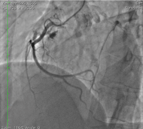
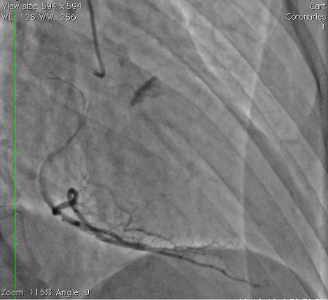
A 32-year-old gentleman was admitted for evaluation of unstable angina. His complete blood count, renal and liver function tests, coagulogram were unremarkable and echocardiography showed normal ejection fraction with no regional wall motion abnormalities. He was planned for coronary angiogram. Right radial artery access was achieved and a 5 Fr Tiger (TerumoTM, Japan) catheter was used. Left coronary artery was essentially normal. The right coronary ostium was hooked, pressure waveform showed no dampening, ostial position was confirmed by a small test injection. However, during the actual contrast injection using manual injection, the catheter super selectively intubated the Conal branch of right coronary artery and 3 ml of dye was injected before stopping the injection. Following this, the distal tip of the Conal artery perforated leading to extravasation of dye into myocardium as shown in Figure 1. His vitals remained stable and an echocardiogram showed no effusion. Few minutes later check angiography showed no further leak from the perforation site and confirmed the persistence of dye staining of the myocardium as shown in Figure 2. Post procedurally he was kept under observation for the next 48 hours and monitored for effusion. His vitals remained stable and no pericardial effusion developed and he was discharged after 3 days.
Discussion
Diagnostic coronary angiogram is a safe procedure with the major complication rate of less than 1%, mortality (<0.1%), myocardial infarction (<0.1%) [1]. Minor complications include vascular complications such as distal embolization, acute thrombosis, bleeding, pseudoaneurysm, arteriovenous fistula, acute kidney injury due to contrast induced nephropathy, allergic reactions to contrast or local anaesthetics, infection and radiation exposure [1,2]. Coronary artery branch perforation is a very rare complication following diagnostic coronary angiogram, although it is not an uncommon complication during angioplasty. The incidence of coronary perforation following angioplasty ranges from 0.2- 0.6% [3], ranging from barely perceptible to severe life-threatening tamponade requiring urgent pericardiocentesis and other interventions like balloon tamponade, deployment of covered stent, use of coil or fat embolization and sometimes emergent or urgent cardiac surgery [4]. The mortality following perforation ranges from 5 to 10 percent [3]. Power Injectors are routinely used when high volume injections are required such as in Ventriculograms, aortograms and run off studies. But greater risk related to the introduction of high pressure in relatively small and delicate vasculature and higher pressures may increase the risk of perforation lead to the use of manual syringe for coronary angiograms.
Coronary perforation following angiography is a very rare complication with only three cases reported in the literature till now. In two of the cases the right coronary artery branch was involved and in one case side branch of the left main coronary artery was involved [5,6]. In our case when a 5 Fr Tiger (TerumoTM, Japan) catheter was used to inject contrast into the right coronary artery, just before injecting the catheter tip accidentally intubated the branch of right coronary artery super selectively leading to the perforation of the tip of the branch, even though the position of the catheter was confirmed prior by a test injection. Perforation of the branch lead to accumulation of the dye in myocardium. An immediate echocardiogram showed no pericardial effusion. In our case the patient has not developed any hemodynamic compromise or effusion so he was kept under conservative management and followed up for development of any pericardial effusion. We conclude that conservative management can help in some cases of perforation during angiography provided the perforation was limited to myocardium and no effusion is noted. We also conclude that pressure tracings should always be monitored during angiography and dye injection to be stopped immediately when there is deep cannulation of the branches.
Conclusion
Coronary angiogram is a relatively safe procedure in current generation. However, complications such as side branch perforation can occur. Pressure tracings should always be monitored during the procedure and dye injection should be stopped when there is deep cannulation of the branches. Conservative management will suffice in a hemodynamically stable patient if no effusion is seen.
- Tavakol M, Ashraf S, Brener SJ (2012) Risks and complications of coronary angiography: a comprehensive review. Glob J Health Sci 4: 65-93. Link: https://bit.ly/2zTJA7Z
- King KM, Ghali WA, Faris PD, Curtis MJ, Galbraith PD, et al. (2004) Sex differences in outcomes after cardiac catheterization: effect modification by treatment strategy and time. JAMA 291: 1220-1225. Link: https://bit.ly/2LMXgVe
- Gruberg L, Pinnow E, Flood R, Bonnet Y, Tebeica M, et al. (2000) Incidence, management, and outcome of coronary artery perforation during percutaneous coronary intervention. Am J Cardiol 86: 680-6822. Link: https://bit.ly/3bRx9qG
- Briguori C, Nishida T, Anzuini A, Di Mario C, Grube E, et al. (2000) Emergency polytetrafluoroethylene-covered stent implantation to treat coronary ruptures. Circulation 102: 3028-3031. Link: https://bit.ly/2Tnq5vi
- Schobel WA, Voelker W, Karsch KR (1995) Perforation of a side branch of the right coronary artery during selective coronary angiography using 5 French Judkins catheters. Cathet Cardiovasc Diagn 36: 156-159. Link: https://bit.ly/2Zpw9rk
- Timurkaynak T, Ciftci H, Cemri M (2001) Coronary artery perforation: a rare complication of coronary angiography. Acta Cardiol .56: 323-325. Link: https://bit.ly/3g8y21o


